What Does Breast Cancer Look Like On Imaging : MRI assessment of breast parenchymal enhancement may ... : Instead, breast skin can become thick, red, and look pitted, like an orange peel.
Dapatkan link
Facebook
X
Pinterest
Email
Aplikasi Lainnya
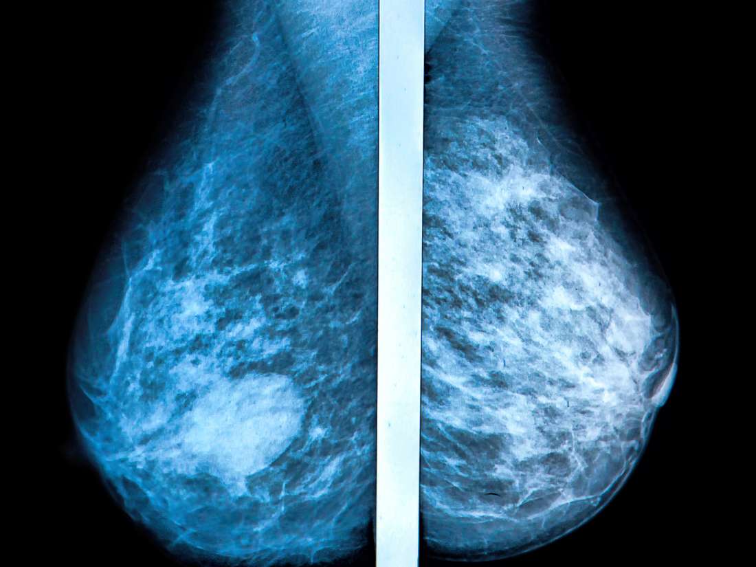
What Does Breast Cancer Look Like On Imaging : MRI assessment of breast parenchymal enhancement may ... : Instead, breast skin can become thick, red, and look pitted, like an orange peel.. A lump or tumor will show up as a focused white area on a mammogram. Inflammatory breast cancer accounts for approximately 5% of all cases of invasive breast cancer in the united states. What does breast cancer look like on a mammogram? Ultrasound is useful for looking at some breast changes, such as lumps (especially those that can be felt but not seen on a mammogram) or changes in women with dense breast tissue. Screening mammograms have been used since the 1980s.
Any area that does not look like normal tissue is a possible cause for concern. What does breast cancer look like on a mammogram? Ductal carcinoma in situ is usually seen as linear microcalcifications (see arrows), which demonstrate linear orientation, as seen in this patient. While many people associate calcifications with breast cancer, there are a number of other potential causes, ranging from benign breast. Rapid onset of symptoms (redness, swelling, warmth, itching, skin thickening) are hallmarks of the disease.
Mammogram images: Normal, abnormal, and breast cancer from cdn-prod.medicalnewstoday.com Magnetic resonance imaging (mri) of the breast — or breast mri — is a test used to detect breast cancer and other abnormalities in the breast. This type of cancer precedes the development of a distinct mass, lump or invasive cancer. A picture is worth a thousand words. Rapid onset of symptoms (redness, swelling, warmth, itching, skin thickening) are hallmarks of the disease. It also can be used to look at a suspicious area that was seen on a mammogram. The dye collection in the breast can also look clumpy or appear in a section of the breast, depending on the involvement of dcis. Mammogram imaging may show skin thickening, but often there is no distinct mass found on physical examination or mammogram. Tumor size is an important factor in breast cancer staging, and it can affect a person's treatment options and outlook.
Breast calcifications are calcium deposits that appear as white dots on a mammogram.
Your doctor will begin to determine this during surgery to remove the cancer and look at one or more of the underarm lymph nodes, which is where breast cancer tends to travel first. What does breast cancer look like on a mammogram? They can vary in type (i.e., size), pattern, and arrangement, and the significance of each of these can vary considerably. Learn about getting a mammogram. The lesions are often rounded tall as broad. A lump or tumor will show up as a focused white area on a mammogram. It can also be used to investigate breast problems, such as a suspicious lump or thickening. That makes it easy to detect abnormalities, which generally show up as white. The doctor reading your mammogram will be looking for different types of breast changes, such as small white spots called calcifications, larger abnormal areas called masses, and other suspicious areas that could be signs of cancer. Screening mammograms have been used since the 1980s. Imaging and lobular breast cancer. Any area that does not look like normal tissue is a possible cause for concern. This type of cancer precedes the development of a distinct mass, lump or invasive cancer.
The doctor reading your mammogram will be looking for different types of breast changes, such as small white spots called calcifications, larger abnormal areas called masses, and other suspicious areas that could be signs of cancer. What does breast cancer look like on a mammogram? Ultrasound is frequently used to evaluate breast abnormalities that are found with screening mammography or diagnostic mammography or during a physician performed clinical breast exam.ultrasound allows significant freedom in obtaining images of the. This is considered an abnormal mammogram, but not necessarily one that's indicative of cancer. Any area that does not look like normal tissue is a possible cause for concern.
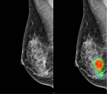
AI-assisted Radiologists Can Detect More Breast Cancer ... from www.itnonline.com While many people associate calcifications with breast cancer, there are a number of other potential causes, ranging from benign breast. The lesions are often rounded tall as broad. A rash isn't the only visual symptom of inflammatory breast cancer. Inflammatory breast cancer accounts for approximately 5% of all cases of invasive breast cancer in the united states. What does breast cancer look like on a mammogram? Ultrasound is frequently used to evaluate breast abnormalities that are found with screening mammography or diagnostic mammography or during a physician performed clinical breast exam.ultrasound allows significant freedom in obtaining images of the. Reiland carries this small rock with her to demonstrate for women what breast cancer feels like. As with all abnormalities seen on breast imaging, the diagnosis of dcis requires a sample of tissue or biopsy.
A breast mri captures multiple images of your breast.
A picture is worth a thousand words. They can vary in type (i.e., size), pattern, and arrangement, and the significance of each of these can vary considerably. In this mammogram image, the breast calcifications are in ductal patterns. Dense breast tissue appears solid. Tumors are likely to be smaller when doctors detect them early, which can. Breast mri images are combined, using a computer, to create detailed pictures. (1) gary ulaner, md, phd, facnm. These images are called mammograms. This is considered an abnormal mammogram, but not necessarily one that's indicative of cancer. Ultrasound is useful for looking at some breast changes, such as lumps (especially those that can be felt but not seen on a mammogram) or changes in women with dense breast tissue. He or she also may order additional blood tests or imaging tests if there is reason to believe the cancer might have spread beyond the breast. A breast mri usually is performed after you have a. It also can be used to look at a suspicious area that was seen on a mammogram.
Inflammatory breast cancer accounts for approximately 5% of all cases of invasive breast cancer in the united states. The most characteristic pathologic feature of tumor emboli within dermal lymphatic vessels is not always detected on skin biopsy 4. Dense breast tissue appears solid. A screening mammogram is performed at regular intervals to check for breast cancer in women who have no signs or symptoms of the disease. The doctor reading your mammogram will be looking for different types of breast changes, such as small white spots called calcifications, larger abnormal areas called masses, and other suspicious areas that could be signs of cancer.
Breast density — MetriTrack from images.squarespace-cdn.com In this mammogram image, the breast calcifications are in ductal patterns. This is considered an abnormal mammogram, but not necessarily one that's indicative of cancer. This type of cancer also changes the appearance of your breasts. What does breast cancer look like on a mammogram? Tumor size is an important factor in breast cancer staging, and it can affect a person's treatment options and outlook. Magnetic resonance imaging (mri) of the breast — or breast mri — is a test used to detect breast cancer and other abnormalities in the breast. What does breast cancer look like on a mammogram? Your doctor will begin to determine this during surgery to remove the cancer and look at one or more of the underarm lymph nodes, which is where breast cancer tends to travel first.
The lesions are often rounded tall as broad.
It also can be used to look at a suspicious area that was seen on a mammogram. As with all abnormalities seen on breast imaging, the diagnosis of dcis requires a sample of tissue or biopsy. Mammogram imaging may show skin thickening, but often there is no distinct mass found on physical examination or mammogram. This type of cancer also changes the appearance of your breasts. A breast mri captures multiple images of your breast. He or she also may order additional blood tests or imaging tests if there is reason to believe the cancer might have spread beyond the breast. A lump or tumor will show up as a focused white area on a mammogram. Ductal carcinoma in situ is usually seen as linear microcalcifications (see arrows), which demonstrate linear orientation, as seen in this patient. These images are called mammograms. While many people associate calcifications with breast cancer, there are a number of other potential causes, ranging from benign breast. A 3d mammogram is used as a breast cancer screening test to look for breast cancer in people with no signs or symptoms of the disease. The lesions are often rounded tall as broad. A diagnostic mammogram is used to check for breast cancer when there is a sign or symptom of disease.
However, shares took a … Find the latest didi global inc. 03.12.2021 · didi global inc (nyse:didi)'s stock surged in u.s. 21, 2021 at 1:47 p.m. Didi global a aktie und aktueller aktienkurs. Didi Delisting Chinese Firm Is Delisting From New York Just Months After Its Disastrous Ipo Cnn from cdn.cnn.com Didi chuxing nyse updated dec 2, 2021 12:59 am. Adrs) | a3ctlg | didi | us23292e1082 (didi) stock quote, history, news and other vital information to help you with your stock trading and investing. Nachrichten zur aktie didi global inc (a) (spons. Didi says it plans to delist from the new york stock exchange immediately. the chinese. However, shares took a … Stock exchanges, a stunning reversal following demands from chinese regulators that had opposed its american listing. 21, 2021 at 1:47 p.m. Stock exchanges, a stunni...
How To Make A Social Security Card : Social Security card caution - YouTube / Looking for duplicate social security card maker online? . The social security card also has a line for the person's signature. We also provide adobe photoshop software with will help you in editing the template. Template social security card usa. Follow the steps below to spot a fake social security card. First, download ss card template psd. The birth is registered and recorded at the belize birth registry; All you need to do is log in to or create your personal my social security account. Easy to customize, layer based.tutorial video you can edit this template and put any : We can create new social security card with your information. Are not requesting a name change or any other change to your card; Father-in-law's first Social Security card - a metal plate ... from i.redd.it ...
How to play backgammon for beginners. Course materials, exam information, and professional development opportunities for ap teachers and coordinators. Detailed information can be found in etsy's cookies & similar technologies policy and. Many of the world's classic board games fit into this category, including: How to play spades with three people. Pin by Adam Wurster on Desktop Setups | Bedroom setup from i.pinimg.com Lewis county noxious weed control. Stepping stones ( great for bodily kinesthetic learners) monopoly jr. Wildlife and fisheries sciences job board a resource provided by the department of rangeland, wildlife and fisheries management. An abstract strategy game is a strategy game in which the theme is not important to the experience of playing. Many of the world's classic board games fit into this category, inc...


Komentar
Posting Komentar